Covid 19 Radiologist Perspective Chest X Ray
Chest x-rays are complex to report accurately. In this article we adopt chest X-ray images instead of computer tomography scans to fine-tune the three proposed classification models.

Imaging In Corona Virus Disease 2019 Covid 19 A Scoping Review European Journal Of Radiology Open
Chest CT and X-ray are key tools in the diagnosis and management of numerous respiratory ailments but their role in the management of COVID-19 has not been considered within the context of many.

Covid 19 radiologist perspective chest x ray. Radiology perspective of coronavirus disease 2019 COVID-19. Clinical team places order for radiology diagnostic imaging study in APEX on COVID-19 confirmed or suspected patient Radiology technologist receives order and brings it to the appropriate radiology service. Lessons from severe acute respiratory syndrome and middle east respiratory syndrome.
As such an important part of managing. The more straightforward descriptions of CT findings can clarify findings on chest radiographs. In our experience baseline CXR had a sensitivity of 681.
In late February researchers at Mount Sinai in New York reported on a retrospective study of chest CTs of 121 symptomatic patients infected with COVID-19. As COVID-19 addresses the epithelial cells lining our airways we use a chest X-ray image in order to analyze the health of a patients lung. Viral pneumonia is often subtle in its radiological appearance.
Coronavirus Disease 2019 COVID -19. Radiology 2020 Jan 31200236 Epub ahead of print. Chest X-ray in new Coronavirus Disease 2019 COVID-19 infection.
Chung M Bernheim A Mei X et al. A new artificial intelligence AI platform developed by Northwestern University researchers can detect COVID-19 in the lungs 10 times faster and a bit more accurately than specialized cardiothoracic radiologists according to a study published today in Radiology. Finally all radiology is a business like any other and we have canceled or deferred almost all elective imaging procedures to help stall the spread of infection and make room for a potential surge of COVID-19 cases the radiologist explained.
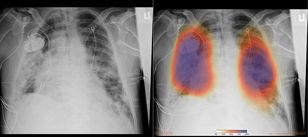
Opacities often have an extensive geographic distribution. In the context of the COVID-19 pandemic rapid triage of cases and exclusion of other pathologies with artificial intelligence AI can assist over-stretched radiology departments. Frequency and Distribution of Chest Radiographic Findings in COVID-19 Positive Patients.
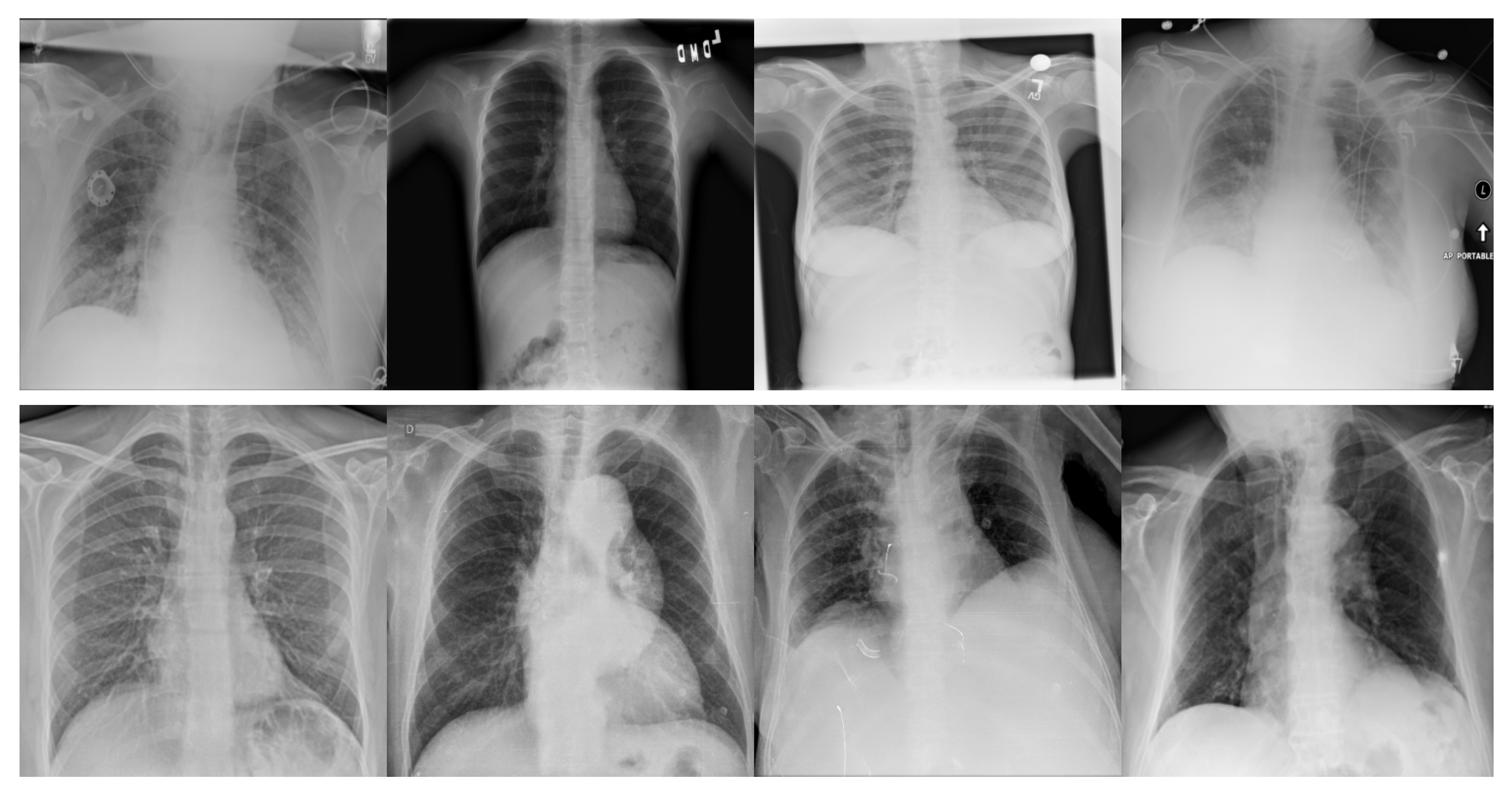
Therefore in the proposed research activity normal pneumonia and Covid-19 chest images have been used and shown in Fig. A Perspective from China. A chest X-ray radiograph is the most commonly ordered imaging study for patients with respiratory complaints.
However many studies are limited by selection bias potential blinding issues and potential confounding of chest CT findings owing to the simultaneous presence of other lung diseases. In this retrospective observational study we enrolled all patients presenting to the emergency department of a Milan-based university hospital from February 24th to April 8th 2020 who underwent nasopharyngeal swab for reverse transcriptase-polymerase. In general we do not recommend follow-up chest X-rays in patients who have had no signs of pneumonia on imaging tests or in whom complete resolution of lung parenchymal involvement was already documented in tests.
A team of LSU Health New Orleans radiologists investigated the usefulness of chest X-rays in COVID-19 and found they could aid in a rapid diagnosis of the disease especially in areas with limited. Because similar pulmonary syndromes have been reported from other strains of the. Coronavirus disease COVID-19 detection in Chest X-Ray images using majority voting based classifier ensemble.
Since the outbreak of the novel coronavirus pulmonary illness coronavirus disease 2019 COVID-19 in China more than 79000 people have contracted the virus worldwide. The most common pathologies are described and the classification of COVID19 appearance in. Chest CT Appearance of COVID-19.
The medical conditions associated with SARS-CoV-2 infections have resulted in a surge in the number of patients at clinics and hospitals leading to a significantly increased strain on healthcare resources. The researchers trained and tested DeepCOVID-XR a machine-learning algorithm that analyzes. Feb 21 2020 Kanne JP.
To this guidance include chest x-rays on inpatients and chest x-rays or CT scans in ED patients. Radiology 2020 Feb 4200230 Epub ahead of. The virus is rapidly spreading with human-to-human transmission despite imposed precautions.
CT Imaging of the 2019 Novel Coronavirus 2019-nCoV Pneumonia. We aim to validate three open-source AI models on an external test. The predominant CT findings of COVID-19 infection are bilateral peripheral and basal predominant ground-glass opacity consolidation or both 6 7.
Wong H Lam H Fong A. To report real-world diagnostic performance of chest x-ray CXR readings during the COVID-19 pandemic. CT Imaging Features of 2019 Novel Coronavirus 2019-nCoV.
This software is by no means a stand-alone solution in the identification of images of COVID-19 from Chest X-rays but can help radiologists and clinicians to achieve a faster and understandable diagnosis using the full potential of Deep Learning without the prerequisite of having to code in any programming language. The world is still struggling in controlling and containing the spread of the COVID-19 pandemic caused by the SARS-CoV-2 virus. Several studies have been published reporting chest CT findings in COVID-19.
Computer Tomography CT X-Ray and Ultrasound US techniques are being applied to scan the patient to determine the presence and severity of Covid-19 34. In particular CXR is a widespread relatively. Chest Imaging Appearance of COVID-19 Infection.
But in patients with severe disease their X-ray readings may resemble pneumonia or acute respiratory distress syndrome ARDS. The expected workflow is as follows. American Journal of Roentgenology.
Around the world radiologists are researching the use of chest CT in COVID-19 cases to help determine the indications of the disease and the role imaging information might play. This investigation includes the scientific work concerning chest radiography chest X-ray - CXR and computed tomography CT in COVID19 patients. In the early stages of COVID-19 a chest X-ray may be read as normal.
In this context thoracic imaging specifically chest X-ray CXR and computed tomography CT is playing an essential role in the management of patients especially those evidencing risk factors from triage phases or moderate to severe COVID-19 signs of pulmonary disease Rubin et al 2020. In COVID-19 CXR shows patchy or diffuse reticular-nodular opacities and consolidation with basal peripheral and bilateral predominance. Findings and correlation with clinical outcome.
Zu ZY Xu PP Chen W et al. Kooraki S Gholamrezanezhad A Reddy S Myers L. Lei J Li J Li X Qi X.
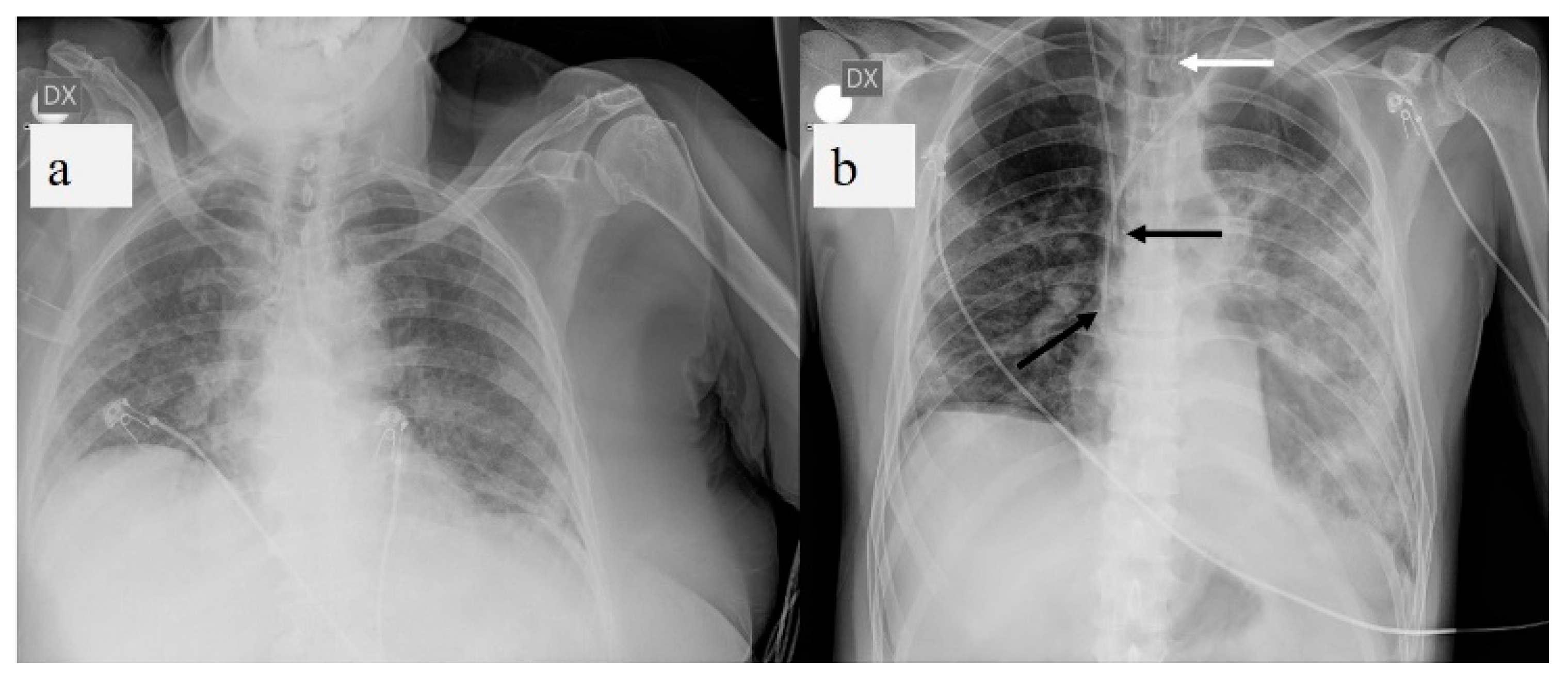
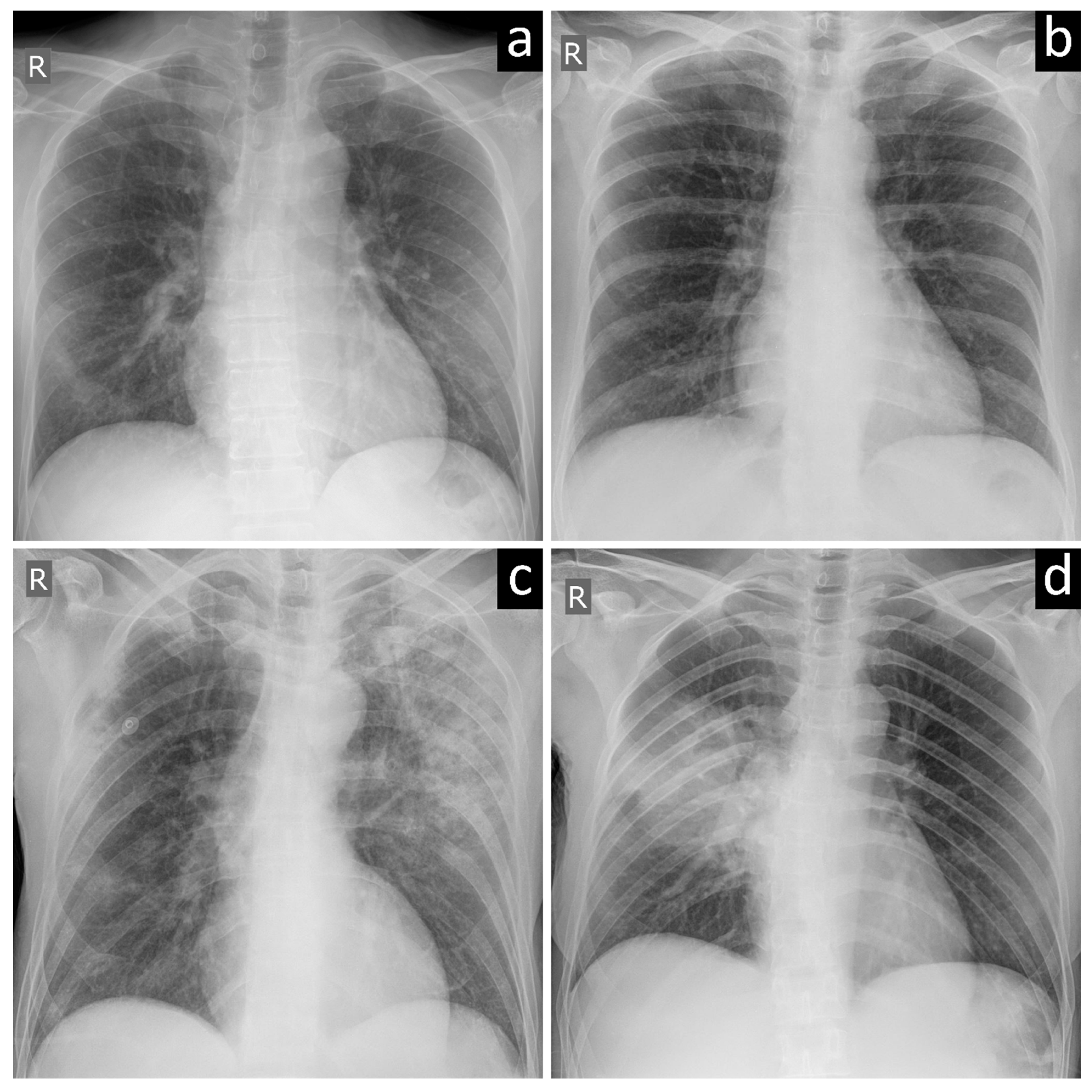
D 58-year-old female post-COVID-19 whose follow-up chest X-ray shows bilateral basal band opacities in relation to laminar atelectasis.
Ai Enables Rapid Covid 19 Lung Imaging Analysis At Ucsd Health Uc It Blog

False Negative Chest X Rays In Patients Affected By Covid 19 Pneumonia And Corresponding Chest Ct Findings Radiography
Diagnostics Free Full Text Fine Tuning Convolutional Neural Networks For Covid 19 Detection From Chest X Ray Images Html
Chest X Ray Findings In 636 Ambulatory Patients With Covid 19 Presenting To An Urgent Care Center A Normal Chest X Ray Is No Guarantee Journal Of Urgent Care Medicine

Chest X Ray In The Covid 19 Pandemic Radiologists Real World Reader Performance European Journal Of Radiology

Imaging Of Covid 19 Pneumonia A Critical Care Perspective Litfl
Diagnostics Free Full Text A Pictorial Review Of The Role Of Imaging In The Detection Management Histopathological Correlations And Complications Of Covid 19 Pneumonia Html
Chest X Ray In The Covid 19 Pandemic Radiologists Real World Reader Performance European Journal Of Radiology
:max_bytes(150000):strip_icc()/covid-19-pneumonia-12-20adbdbe7ee54f7784689c3b1ede2d1c.jpg)
Covid 19 Coronavirus Diagnosis Chest X Ray And Ct Scan

X Ray Imaging For Covid 19 Patients

Chest X Ray Or Ct For Covid 19 Pneumonia Comparative Study In A Simulated Triage Setting European Respiratory Society

Covid 19 In The Radiology Department What Radiographers Need To Know Radiography

Diagnostic Impact Of Bedside Chest X Ray Features Of 2019 Novel Coronavirus In The Routine Admission At The Emergency Department Case Series From Lombardy Region European Journal Of Radiology
Diagnostics Free Full Text Artificial Intelligence Applied To Chest X Ray For Differential Diagnosis Of Covid 19 Pneumonia Html

X Ray Imaging For Covid 19 Patients

Diagnostic Imaging Of Covid 19 Patients Tidsskrift For Den Norske Legeforening

Utility Of Screening Chest Radiographs In Patients With Asymptomatic Or Minimally Symptomatic Covid 19 In Singapore Radiology

Chest X Ray In The Covid 19 Pandemic Radiologists Real World Reader Performance European Journal Of Radiology

X Ray Imaging For Covid 19 Patients
Post a Comment for "Covid 19 Radiologist Perspective Chest X Ray"